
Vascular
Vascular Neuropathology
What You'll Learn
- The pathology and timeline of ischemic infarction and global hypoxic-ischemic injury
- Hemorrhagic stroke by location — deep (hypertensive) vs lobar (amyloid) — and subarachnoid hemorrhage / aneurysms
- Vascular malformations — AVM, cavernoma, developmental venous anomaly, capillary telangiectasia
- Small-vessel disease — lacunes, cerebral amyloid angiopathy, CADASIL — plus CNS vasculitis and venous thrombosis
This is the neuropathology view — gross, histology and stains for each entity. Clinical management lives in the Vascular clinical notes.
🔍 High-Yield Pearls
- Infarct timeline: red neurons (12–24 h) → neutrophils (1–2 d) → foamy Gitter cells (days–weeks) → cystic cavity + gliosis (weeks–months). The brain uniquely undergoes liquefactive necrosis.
- Location tells the cause: deep (basal ganglia/thalamus/pons/cerebellum) = hypertensive; lobar in the elderly = cerebral amyloid angiopathy.
- Saccular (berry) aneurysms at circle-of-Willis branch points (ACOM > PCOM > MCA bifurcation) cause SAH; delayed vasospasm peaks days 4–14.
- Cavernoma = back-to-back sinusoidal vessels with no intervening brain (angiographically occult, “popcorn” on MRI); AVM = a nidus of arteries feeding arterialized veins with no capillary bed.
- CADASIL = NOTCH3; granular osmiophilic material (GOM) on EM — migraine + recurrent subcortical strokes + dementia.
Ischemic Infarction (Arterial)
Mechanism: thrombotic (atherosclerotic plaque rupture, in-situ) or embolic (cardiac/artery-to-artery; often to the MCA territory, hemorrhagic on reperfusion). Result is liquefactive necrosis in a vascular territory.
Histologic evolution
| Time | On the slide | Cells |
|---|---|---|
| 0–12 h | No H&E change (cytotoxic edema only) | — |
| 12–24 h | Red (eosinophilic) neurons; pallor | Dying neurons |
| 1–3 d | Neutrophil infiltration; tissue necrosis | Neutrophils |
| 3–7 d | Macrophages digest myelin/debris | Gitter cells (foamy macrophages) |
| 1–3 wk | Liquefaction; reactive gliosis at rim; neovascularization | Macrophages, reactive astrocytes |
| >1 mo | Cystic cavity walled by astrogliosis | Astrocytes |
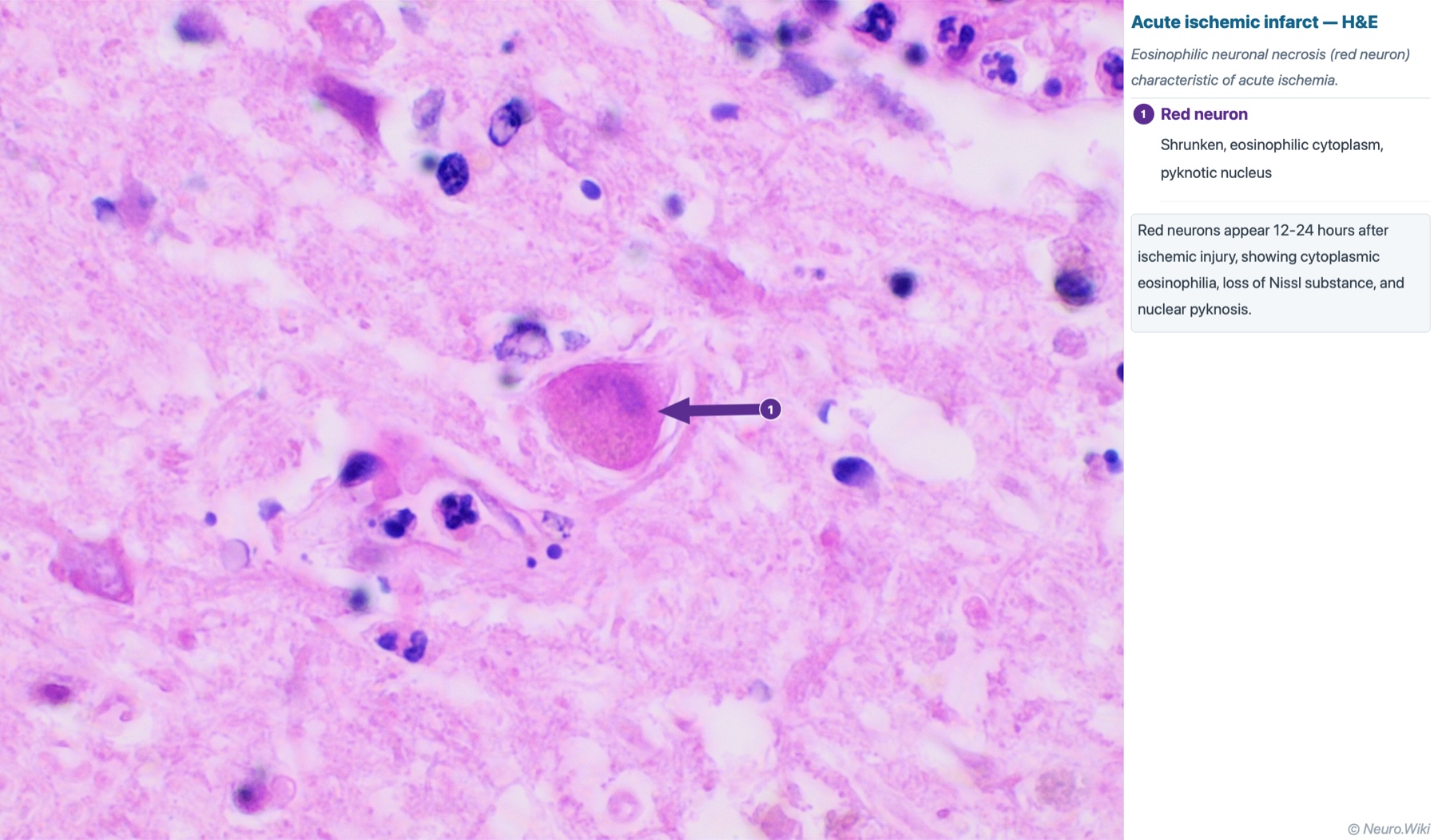
Hemorrhagic transformation: reperfusion into damaged, leaky vessels (embolic infarcts, post-thrombolysis). Remote infarct: a gliotic-walled cystic cavity (cystic encephalomalacia).
Global Hypoxic-Ischemic Injury
Diffuse hypoperfusion/hypoxia (cardiac arrest) injures the most metabolically demanding, selectively vulnerable zones:
- Hippocampus CA1 (Sommer sector) — the most vulnerable neurons.
- Cerebellar Purkinje cells and neocortical layers 3 & 5 (→ laminar/pseudolaminar necrosis).
- Watershed (border-zone) infarcts between major arterial territories (e.g., ACA-MCA), classically after systemic hypotension.
Intracerebral Hemorrhage
| Type | Location | Vessel pathology / stain |
|---|---|---|
| Hypertensive | Deep: putamen/external capsule > thalamus > pons > cerebellum | Lipohyalinosis / fibrinoid necrosis of penetrating arterioles (classic Charcot-Bouchard microaneurysms) |
| Cerebral amyloid angiopathy | Lobar (cortical/subcortical), elderly/normotensive; recurrent | Aβ in cortical/leptomeningeal vessel walls; Congo red apple-green birefringence |
Subarachnoid Hemorrhage & Aneurysms
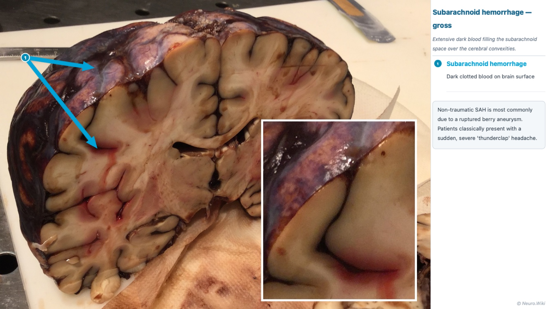
Saccular (berry) aneurysms form at circle-of-Willis branch points: anterior communicating (most common) > posterior communicating > MCA bifurcation. Rupture → blood fills the subarachnoid space / basal cisterns.
- Associations: ADPKD, Ehlers-Danlos type IV, coarctation, smoking/hypertension, fibromuscular dysplasia.
- Complications: re-bleed (early), vasospasm → delayed ischemia (days 4–14), communicating hydrocephalus, seizures.
- Other aneurysms: fusiform (atherosclerotic, basilar), mycotic (infective endocarditis, distal), Charcot-Bouchard (microaneurysm → ICH, not SAH).
Vascular Malformations
| Malformation | Pathology | Key point |
|---|---|---|
| Arteriovenous malformation (AVM) | Tangled nidus of abnormal arteries and arterialized (thick, hyalinized) veins with no intervening capillary bed; gliotic brain between vessels | Highest bleed risk; supratentorial; seizures/hemorrhage in young adults |
| Cavernous malformation (cavernoma) | Well-circumscribed cluster of back-to-back thin sinusoidal vessels with no intervening brain; surrounding hemosiderin/gliosis | Angiographically occult; “popcorn/mulberry” on MRI; low-flow; familial (CCM genes) |
| Developmental venous anomaly (DVA) | Radially arranged dilated veins draining to a single collector (caput medusae) through normal brain | Benign, usually incidental; often accompanies a cavernoma |
| Capillary telangiectasia | Dilated thin capillaries separated by normal brain, typically pons | Benign, incidental |
Small-Vessel Disease
| Entity | Pathology / stain | Clinical clue |
|---|---|---|
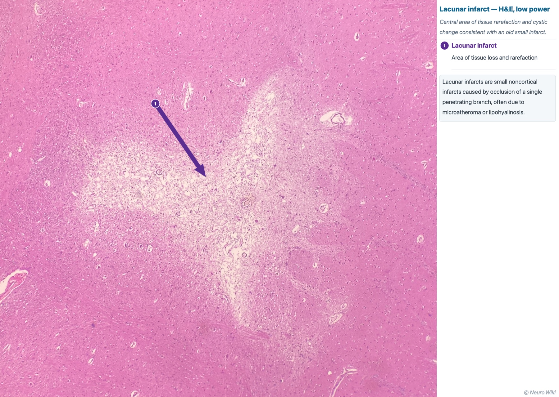
| Lacunar infarct | Small (<15 mm) cavity + macrophages from lipohyalinosis of a deep penetrator | Pure motor (internal capsule/pons) or pure sensory (thalamus) |
| Cerebral amyloid angiopathy | Aβ in cortical/leptomeningeal vessels; Congo red apple-green | Recurrent lobar hemorrhage, elderly |
| CADASIL | Media thickening; granular osmiophilic material (GOM) on EM; NOTCH3 | Migraine + subcortical strokes + dementia; AD inheritance |
| Hypertensive arteriolosclerosis (Binswanger) | Hyaline arteriolosclerosis → diffuse subcortical white-matter rarefaction | Vascular cognitive impairment, gait |
CNS Vasculitis & Venous Thrombosis
- Primary angiitis of the CNS (PACNS): transmural inflammation ± granulomas and fibrinoid necrosis of small/medium leptomeningeal and parenchymal vessels → multifocal ischemia; biopsy (leptomeninges + cortex) confirms.
- Secondary vasculitis: infection (VZV, TB, syphilis), systemic (PAN, GPA, SLE), drug (cocaine/amphetamine).
- Cerebral venous / dural sinus thrombosis: hemorrhagic venous infarct not respecting an arterial territory (often bilateral parasagittal with superior sagittal sinus thrombosis); risk = hypercoagulable states, pregnancy/puerperium, OCPs, dehydration, local infection.
High-Yield Facts
| Finding | Buzzword / association |
|---|---|
| Red neurons | Acute ischemia, 12–24 h; CA1 & Purkinje most vulnerable |
| Deep ganglionic hemorrhage | Hypertensive; Charcot-Bouchard / lipohyalinosis |
| Lobar hemorrhage, elderly | Cerebral amyloid angiopathy (Congo red) |
| Berry aneurysm, ACOM | SAH; vasospasm days 4–14; ADPKD/EDS |
| No intervening brain between vessels | Cavernoma (occult) or AVM (arterialized veins) |
| Caput medusae | Developmental venous anomaly (benign) |
| GOM on EM / NOTCH3 | CADASIL |
| Bilateral parasagittal hemorrhagic infarct | Superior sagittal sinus thrombosis |
References
- Love S, Budka H, Ironside JW, Perry A. Greenfield's Neuropathology. 9th ed. CRC Press; 2015.
- Ellison D, Love S, et al. Neuropathology. 3rd ed. Mosby; 2013.
Continue reading — sign in
The full note has more clinical pearls, tables, and board-focused tips. Free account, no fee.